Discover i-SCAN.
Get more diagnostic information and gain greater clinical confidence with i-SCAN Advanced Imaging, exclusively from PENTAX Medical.
- I-SCAN highlights vascularity and surface anomalies, assisting in visibility of pathology
- i-SCAN highlights edges of lesions assisting in surgical planning
- i-SCAN can be used concurrent with stroboscopy for enhanced visualization with motion
Case Descriptions.
Case 1: Is an exam of a 23-year-old male with Recurrent Respiratory Papillomatosis. When utilizing i-SCAN the vascularity and surface anomalies are highlighted, providing additional information for diagnosis and treatment planning.
Case 2: Depicts early esophageal neoplasia arising in a patient with Barrett`s esophagus. When combining i-SCAN imaging with acetic acid chromoendoscopy the edges of the lesion becomes more apparent, as the abnormal irregular mucosal architecture is enhanced.
Case 3: Is an exam of a 50-year-old male who was found to have leukoplakia on his left vocal fold. This exam demonstrates how i-SCAN can be used concurrent with stroboscopy, for enhanced visualization with motion.
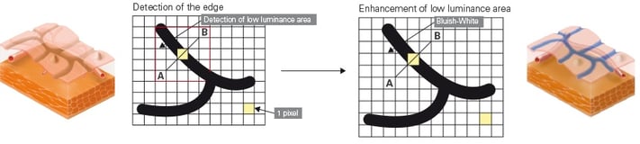
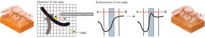
How it works.
- i-SCAN is a software-based image processing technology that enhances digital images for display and visualization.
- i-SCAN modes combine image processing algorithms for Surface Enhancement (SE), Contrast Enhancement (CE), and Tone Enhancement (TE) to provide a digitally enhanced view of mucosal surface texture, contour, and blood vessels.
- Contrast Enhancement (CE) CE provides a sharper distinction between edges and contrasting color by increasing the appearance of light-dark transistions.
- Surface Enhancement (SE) provides a more three-dimensional effect to an image by adjusting luminous contrast between pixels.
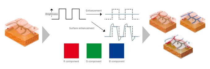
- Tone Enhancement (TE) digital tone filters to apply color contrast between the Red, Green and Blue channels of the image.